Hi everyone, gwen here. I am going to share with you about MTT assay. This assay is done to determine the potency of the drug and proteins used for cancer therapy. Each treatment can consist of the drug with the protein or just the drug or protein alone.
The first step to this assay would be to seed the desired cells into 96-well plates. The number of plates needed would depend on the number of treatments planned. The cells would then be incubated and treated with treatments the next day. Each plate will have 12 columns altogether. 2 columns are reserved as controls and the cells in the remaining 10 columns of cells will be treated with the treatments.
After overnight incubation, the MTT (3-(4,5-Dimethylthiazol-2-yl)-2,5-diphenyltetrazolium bromide, a tetrazole) solution is added to the cells after removing the used media/treatments. The plates are then incubated for another 3-5 hours. During this time, the MTT forms purple formazan from the living cells. After the incubation, the remaining MTT solution would be removed. Care must be taken so as not to pipette the purple formazan out, as it will affect the viability results.DMSO is then added to each well of the plate so as to dissolve the purple formazon, then the absorbance level can be read and the viability of the cells determined. Viability is calculated by dividing the absorbance level of the treated cells with that of the control and a percentage is derived.
Wednesday, August 26, 2009
Sunday, August 23, 2009
Laboratory techniques - surgical castration
Hello everyone! im back to share with you all about surgical castration of rats!
Castrated rats would have testosterone levels near null levels because of the removal of the testes. Therefore, surgical castration of rats is done in my lab to test if our drugs have an effect on testosterone levels in them.
Removal of testes from a rat is a very painful procedure, as it concerns one of the most sensitive organs of the rat. Therefore, these rats have to be anaesthetized and given pain-killers before starting the operation so that it would feel the least pain during removal.
When the rat is fully anaesthetized, we would clean the genital area of the rat with iodine to make sure that it is sterile. Then we make a small slit of about 1cm in the skin at the tip of the scrotum of the rat. Then using forceps, we remove the connective tissues around the sacs so as to isolate the scrotum sacs. Then we suture the top of the scrotum sac so as to prevent blood flow into the testes and then using a sharp scissors, we cut off the whole sac. This procedure is repeated for the other scrotum sac.
When both testes are removed, we check for any signs of bleeding. These openings would then be sewn together to prevent haemorrhage. The inner muscular walls would then be sewn together followed by the skin area. Suture is done discontinuously so that when the rat bites on one of the knots, only one suture would fall out and not the whole line of suture.
After this, the area would be cleaned again with iodine and antibacterial medication is applied on the area of surgery. Antibiotics would also be given to the rat to prevent bacterial infections from occuring.
The rats would be observed until they recover from the anaesthesia. The rats would be given pain-killers, antibiotics and antibacterial medication for the next three days. Overall, the rats for my project that have undergone castrations are recovering well and is slowly recovering activity.
Ok, thats all folks.
will update again soon!
Renee
0703634F
TG02
Monday, August 10, 2009
General Chemistry
It’s my turn to blog again after 5 weeks (: I’ll be sharing about the general chemistry section this time. I did specimen processing at this section so my good friends are Cobas, MPA and SWA. I also need to do basic housekeeping like cleaning the work area with 0.1 hypochlorite and refill consumables like hitachi cups daily. When I receive the blood and urine samples, I must first check that the name on the label and the tube matches before pasting the labels on the tubes. Before samples are processed, I also need to check if there is sufficient volume (at least half the tube is filled), have tests other than biochemistry and whether is it to be loaded on Cobas or SWA. When there is insufficient volume or needs to be loaded on Cobas, I will need to spin it offline (centrifuge manually). If not, I can just load the samples into MPA directly.
I will be sharing about my good friend, MPA, this week. MPA refers to Roche Hitachi Modular Pre-Analytics Standard B Plus. This piece of equipment helps to minimize our exposure to the blood samples. Sample tubes must be racked according to the tube height before loading into MPA. If different heights are placed together in a rack, the alarm will sound. There will be an alarm code and I can check on the chemistry computer to check the reason for the alarm. Samples are identified by LIS generated accession labels. Erroneous labels, labels that cannot be read or labels that are stick in such a way that the machine cannot rotate the tubes, the rack of tubes will be ejected via a rejection buffer for the operator to retrieve. Racks are placed in the Input Buffer Module (IBM) and are immediately transported to the Automatic Centrifuge Unit (ACU), which spins at 1800g for 5minutes or to a pre-centrifuge holding area if the centrifuge is full. This holding area also facilitates the ability for a subsequent urgent request to be inserted into the next available centrifugation cycle. After which, the racks move to the Destopper Module (DSP) to have the caps removed before samples proceed to the Online Aliquoter Module (AQN). At AQN, individual daughter aliquots are made for clinical chemistry, immunoassay, offline testing or archival. Daughter tubes for offline testing are labeled by an online labeler at the Barcode Labeler Module (BCL) and recapped if necessary (e.g. HIV specimens) at the Restopper Module (RSP) or recapped manually using cheap green caps. Before the samples enter BCC, the AQN will check if there is sufficient serum and whether there is any clot. Should a sample have any of those conditions, the sera will be returned to the parent tube and ejected for manual manipulation. For samples clear of those conditions, they will proceed from AQN to BCC to RSP and finally archived in the Flexible Sample Sorter Module (FSS). The sera will then continue on into the SWA for testing or taken out for Cobas testing. Only for MPA, the racks used cannot be reused until data clean is done by the night staff.
I can't find any good pictures to post so here's the link (http://rochediagnostics.ca/lab/solutions/modular-pap.php)for a clearer understanding with labelled picture. Hope it helps(:
Goh Michelle
0703478H
TG02
Wednesday, August 5, 2009
Immulite Analyzer- Laboratory technique
Hello everybody! Its me, zhang’e!
Today, I’m going to explain to you all about an analyzer called IMMULITE, its principle and application. =D
Applications
This analyzer can carry out different tests at the same time. For examples, cortisol test, progesterone test, estradiol test, testosterone test. (These are just a few examples). The analyzer is able to measure the concentration of a compound of interest inside a sample. It is able to provide us a preliminary diagnosis or value to check for any abnormalities. (E.g. you can measure progesterone concentration in patients’ serum to check for their reproduction function or measure the insulin concentration to determine whether the person has diabetes). It is not a confirmatory test.
The test you want to carry out depends on the reagents that you have. For example, I had carried out cortisol test using this analyzer. So, I was given a box containing all the reagents that I needed for cortisol test. (The box is specifically for cortisol test). Different tests have their own boxes.
In each box, there is:
1. Test units (Barcode-labelled unit that contains one bead coated with antibody)
• For cortisol test: Cortisol test unit is used. It contains one bead coated with polyclonal rabbit anti-cortisol.
2. Reagent Wedge (Barcode-labelled unit that contains 7.5ml of enzyme conjugated to compound of interest)
• For cortisol test: Cortisol reagent wedge is used. It contains 7.5ml of alkaline phosphatase (bovine calf intestine) conjugated to cortisol)
• This is the labeled antigens
3. Adjustors (Two vials of adjustors are provided for each test, one high and one low adjustors, each contains 3ml of compound of interest)
• For cortisol test: Cortisol adjustors are used. Each contains 3ml of cortisol.
• Adjustor is like a calibrator which calibrates and ensures that the analyzer is functioning well before we carry out any test. Low adjustor will yield low level of result and high adjustor will yield high level of result.
4. Sample Diluent (One vial contains 25ml of processed human serum with the absence of compound of interest)
• For cortisol test: Cortisol sample diluent is used. It contains 25ml of processed cortisol free human serum. It is used to dilute the concentrated sample.
5. Chemiluminescent Substrate (One vial which contains the substrate for reacting with the enzyme to emit light)
6. Sample Cup Holder (Each cup holder has a labeled number and can be re-used after each test).
• Sample cup holder is the same for all the tests. There is no specific sample cup holder for each test.
7. Sample Cup (Sample is loaded onto the sample cup)
• Sample cup is thrown away after each use.
Principle
Immulite/Immulite 1000 analyzer is an automated machine that is connected to a computer. It performs a solid-phase, competitive chemiluminescent enzyme immunoassay. It involves a competition binding between the labeled antigens and non-labeled antigens to the limited amount of antibody binding sites. (This is somehow similar to RIA).
In cortisol test, alkaline phosphatase (enzyme) is used to label the cortisol (antigen). The labeling is already done and provided in cortisol reagent wedge. Sample with unknown concentration of cortisol is not labeled. After calibrating the analyzer using the adjustors, the wedge is loaded onto the analyzer. At least 100ul of sample is added to the sample cup (if is less than 100ul, the level in the sample cup is too low thus the probe is not able to detect or absorb the sample) and the sample cup is placed onto the sample cup holder. The cup holder is then loaded onto the analyzer. Each sample is loaded together with one test unit (antibody). Chemiluminescent substrate is also loaded. All the information is keyed into the computer (such as the test that you are going to run, the sample cup holder’s number, the name of sample, etc) and the test started to run. The analyzer will mix the sample, labeled cortisol in wedge and antibody (test unit) together. After mixing, it will add the substrate to chemically react with the enzyme to produce light. The light is captured and measured. The amount of bound-labelled antigen is determined and is inversely proportional to the amount of non-labelled antigen. The overall concentration of cortisol in the sample is calculated and generated.
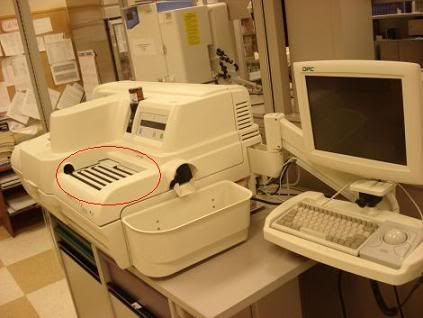
Immulite Analyzer.
Retrieved on August 2, 2009 from website http://www.dotmed.com/images/listingpics/301629.jpg
This is the analyzer. This is when the analyzer is not in use, the cover is close. When it is in use, the whole cover is open.
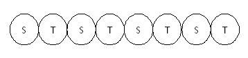
The circle area is where the sample cup holder with the sample cup is loaded at. Each sample is loaded together with one test unit (antibody). An example is shown below. (S=Sample, T=test unit)
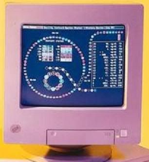
Screen of the computer.
Retrieved on August 2, 2009 from website http://www.vghks.gov.tw/air/images/immulite-4.jpg
This is the screen on the computer that will show you the position of your sample. It takes 40 minutes to complete each run (complete the cycle). The small circle is where the wedges are loaded.
The end! =D
Zhang’e
0704086H
TG02
Today, I’m going to explain to you all about an analyzer called IMMULITE, its principle and application. =D
Applications
This analyzer can carry out different tests at the same time. For examples, cortisol test, progesterone test, estradiol test, testosterone test. (These are just a few examples). The analyzer is able to measure the concentration of a compound of interest inside a sample. It is able to provide us a preliminary diagnosis or value to check for any abnormalities. (E.g. you can measure progesterone concentration in patients’ serum to check for their reproduction function or measure the insulin concentration to determine whether the person has diabetes). It is not a confirmatory test.
The test you want to carry out depends on the reagents that you have. For example, I had carried out cortisol test using this analyzer. So, I was given a box containing all the reagents that I needed for cortisol test. (The box is specifically for cortisol test). Different tests have their own boxes.
In each box, there is:
1. Test units (Barcode-labelled unit that contains one bead coated with antibody)
• For cortisol test: Cortisol test unit is used. It contains one bead coated with polyclonal rabbit anti-cortisol.
2. Reagent Wedge (Barcode-labelled unit that contains 7.5ml of enzyme conjugated to compound of interest)
• For cortisol test: Cortisol reagent wedge is used. It contains 7.5ml of alkaline phosphatase (bovine calf intestine) conjugated to cortisol)
• This is the labeled antigens
3. Adjustors (Two vials of adjustors are provided for each test, one high and one low adjustors, each contains 3ml of compound of interest)
• For cortisol test: Cortisol adjustors are used. Each contains 3ml of cortisol.
• Adjustor is like a calibrator which calibrates and ensures that the analyzer is functioning well before we carry out any test. Low adjustor will yield low level of result and high adjustor will yield high level of result.
4. Sample Diluent (One vial contains 25ml of processed human serum with the absence of compound of interest)
• For cortisol test: Cortisol sample diluent is used. It contains 25ml of processed cortisol free human serum. It is used to dilute the concentrated sample.
5. Chemiluminescent Substrate (One vial which contains the substrate for reacting with the enzyme to emit light)
6. Sample Cup Holder (Each cup holder has a labeled number and can be re-used after each test).
• Sample cup holder is the same for all the tests. There is no specific sample cup holder for each test.
7. Sample Cup (Sample is loaded onto the sample cup)
• Sample cup is thrown away after each use.
Principle
Immulite/Immulite 1000 analyzer is an automated machine that is connected to a computer. It performs a solid-phase, competitive chemiluminescent enzyme immunoassay. It involves a competition binding between the labeled antigens and non-labeled antigens to the limited amount of antibody binding sites. (This is somehow similar to RIA).
In cortisol test, alkaline phosphatase (enzyme) is used to label the cortisol (antigen). The labeling is already done and provided in cortisol reagent wedge. Sample with unknown concentration of cortisol is not labeled. After calibrating the analyzer using the adjustors, the wedge is loaded onto the analyzer. At least 100ul of sample is added to the sample cup (if is less than 100ul, the level in the sample cup is too low thus the probe is not able to detect or absorb the sample) and the sample cup is placed onto the sample cup holder. The cup holder is then loaded onto the analyzer. Each sample is loaded together with one test unit (antibody). Chemiluminescent substrate is also loaded. All the information is keyed into the computer (such as the test that you are going to run, the sample cup holder’s number, the name of sample, etc) and the test started to run. The analyzer will mix the sample, labeled cortisol in wedge and antibody (test unit) together. After mixing, it will add the substrate to chemically react with the enzyme to produce light. The light is captured and measured. The amount of bound-labelled antigen is determined and is inversely proportional to the amount of non-labelled antigen. The overall concentration of cortisol in the sample is calculated and generated.

Immulite Analyzer.
Retrieved on August 2, 2009 from website http://www.dotmed.com/images/listingpics/301629.jpg
This is the analyzer. This is when the analyzer is not in use, the cover is close. When it is in use, the whole cover is open.
The circle area is where the sample cup holder with the sample cup is loaded at. Each sample is loaded together with one test unit (antibody). An example is shown below. (S=Sample, T=test unit)


Screen of the computer.
Retrieved on August 2, 2009 from website http://www.vghks.gov.tw/air/images/immulite-4.jpg
This is the screen on the computer that will show you the position of your sample. It takes 40 minutes to complete each run (complete the cycle). The small circle is where the wedges are loaded.
The end! =D
Zhang’e
0704086H
TG02
Subscribe to:
Posts (Atom)